Specialist Level Diagnostics
Our goal: To help expand the services provided by veterinary practices to further facilitate efficient diagnosis and treatment of patients in one location. We have found that this partnership is beneficial for both patient care and practice health.
Our Goal
For all of our diagnostic services, a preliminary report on paper will be generated at the time of the procedure and handed to the veterinarian in charge of the case. Later the same day we will send through a thorough comprehensive digital report for your patient records.
For procedures requiring assistance, we kindly ask that one of your members of staff be available to assist for the duration of the procedure.
Abdominal Ultrasound
Abdom
Our certified specialists and highly skilled veterinary sonographers will be using the GE Vivid iq mobile, state of the art ultrasound machine with a full complement of transducers. This test will provide insight into the underlying disease process, enabling a diagnosis and formation of a tailored treatment plan. Abdominal ultrasound provides clear and safe guidance for sampling of fluid or organs, mitigating the risk of inadvertent perforation or haemorrhage.
Echocardiography
Echocardiography is a complex, non-invasive and accurate method to diagnose an array of congenital and acquired cardiac and pericardial diseases.
Our certified specialists and highly skilled veterinary sonographers will be using the GE Vivid iq mobile, state of the art ultrasound machine with a full complement of transducers. This test will provide insight into the function and structure of the heart. Through echocardiography, we can assess the size of the chambers, wall thickness, valvular motion and myocardial contractility. Our equipment permits use of B-mode, M-mode, colour flow Doppler including pulsed and continuous wave forms and the latest tissue Doppler imaging.
Thoracic Ultrasound
Thoracic ultrasound is an excellent test to complement thoracic radiographs.
Our certified specialists and highly skilled veterinary sonographers will be using the GE Vivid iq mobile, state of the art ultrasound machine with a full complement of transducers. This test will provide insight into the disease processes involving the mediastinum, lungs, heart and pleural space. Thoracic ultrasound provides a platform for sample collection and enables visualization of vital structures to reduce complications such as haemorrhage or pneumothorax.
Pregnancy Ultrasound
Pregnancy Ultrasound is very sensitive for the diagnosis of pregnancy. Although pregnancy can be diagnosed as early as 17-19 days in dogs and 11-14 days in cats, we recommend scanning at 21-25 days to ensure accuracy.
Our certified specialists and highly skilled veterinary sonographers will be using the GE Vivid iq mobile, state of the art ultrasound machine with a full complement of transducers. This test will provide insight into the fetal viability and gestational age. Ultrasound generally is not an accurate method of determining litter size. Please note that we will always do our best to estimate fetal numbers but cannot guarantee exact numbers.
Neck Ultrasound
Neck ultrasound provides detailed information about the complex endocrine organs of the thyroid and parathyroid glands.
Our certified specialists and highly skilled veterinary sonographers will be using the GE Vivid iq mobile, state of the art ultrasound machine with a full complement of transducers. This test will provide insight into the disease processes involving these glands. Cervical ultrasound aids in the diagnosis of feline hyperthyroidism, canine hypothyroidism, thyroid and parathyroid neoplasia. This procedure requires heavy sedation or general anaesthesia to enable patient compliance and positioning in dorsal recumbency.
Musculoskeletal (MSK) Ultrasound
MSK ultrasound is an invaluable, non-invasive and efficient procedure to thoroughly examine musculature, ligaments, tendons and joints.
Our certified specialists and highly skilled veterinary sonographers will be using the GE Vivid iq mobile, state of the art ultrasound machine with a full complement of transducers. This test will provide insight into the underlying disease process, enabling a diagnosis and formation of a tailored treatment plan.
Endoscopy
** All endoscopy procedures include sample collection
- Endoscopy equipment
- Upper GI endoscopy (Gastroscopy)
Lower GI endoscopy (Colonoscopy) - Combined upper and lower GI
endoscopy - Rhinoscopy
- Otoscopy and myringotomy
- Bronchoscopy and BAL
- Vaginoscopy/Urethroscopy/Cystoscopy
(female dogs only) - Stricture balloon dilation
- Electrosurgical snare polypectomy
- Endoscopically assisted gastropexy
(requires surgeon to complete procedure) - Endoscopy assisted Oesophageal or
Gastric foreign body retrieval
Endoscopy Equipment
We come equipped with a full suite of brand new Karl Storz Veterinary specific endoscopes. This is the latest in state of the art veterinary endoscopy which enables us to perform endoscopy procedures on small and large patients.
We have all of the equipment to perform endoscopy of the ear canals, nasal passages, large airways, urinary tract of females, upper and lower gastrointestinal tract. We also provide all of the necessary ancillary equipment to obtain biopsies, do washes or soaks, dilate strictures and remove foreign bodies.
Endoscopically Assisted Gastropexy
Endoscopically assisted gastropexy is a recent advancement in prophylactic gastropexy that has the added benefits of being minimally invasive and a very short procedure time. This collaborative procedure involves the expertise of an endoscopist with a surgeon. The endoscope is placed into the pyloric antrum and the stomach insufflated. The surgeon then passes two stay sutures into the stomach to secure it to the body wall. The patient then undergoes a minimally invasive procedure which involves dissecting down to the gastric submucosa. The stomach seromuscular layer is then sutured to the peritoneum and transverse abdominal muscle.

Electrosurgical snare polypectomy
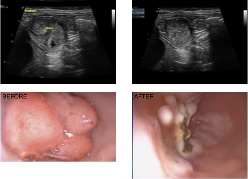
For polyps and tumours located within the GIT, using endoscopic assistance, electrosurgical snare polypectomy can be performed and is an effective minimally invasive non-surgical method to remove solid lesions from within the GIT. A combination of cutting and electrocautery is used to safely remove the lesion with no bleeding.
Gastroscopy
Gastroscopy is a procedure whereby a flexible long camera is placed through the animal’s mouth, down the oesophagus, into the stomach and duodenum. performed under general anaesthesia. Our specialists at Insight use state of the art Karl Storz endoscopy equipment. The endoscope is used to inspect the lining of the stomach and duodenum and to collect biopsy samples to investigate underlying problems.
Common reasons to perform this procedure include vomiting, inappetance, regurgitation etc. And common conditions encountered include Helicbacter infection and Inflammatory Bowel Disease.
Colonoscopy
Colonoscopy is a procedure whereby a flexible long camera is placed through the animal’s rectum and into the large intestine (colon), performed under general anaesthesia. This is done after a gentle warm water enema to clear out faeces. The endoscope is used to inspect the lining and to collect biopsy samples to investigate underlying problems.
Common conditions encountered include Inflammatory Bowel Disease and Histiocytic Ulcerative Coliits.
Cystoscopy
Cystoscopy is a procedure performed under general anaesthesia whereby a rigid camera is placed inside the urethra and extended into the bladder The endoscope is used to inspect the lining and to collect biopsy samples. Our specialists at Insight use state of the art Karl Storz endoscopy equipment.
Common reasons to perform this procedure include recurrent urinary tract infections, tumours and polyps.
Stricture balloon dilation
Strictures are circumferential narrowings commonly encountered in the oesophagus or colon. These can be dilated under general anaesthesia using a special balloon under endoscopic visualisation to allow a wider lumen and easier passage of food or faeces.
Rhinoscopy
Rhinoscopy is a procedure whereby a rigid and flexible camera is placed inside the animal’s nose under general anaesthesia. The endoscope is used to inspect the lining and to collect biopsy samples or remove foreign objects. Our specialists at Insight use state of the art Karl Storz endoscopy equipment.
Common reasons to perform this procedure include mucoid or bloody diarrhoea, straining to defecate etc. Common reasons to perform this procedure include sneezing, facial discomfort and nasal discharge. Comm conditions encountered include inflammatory disease, tumours, foreign bodies and fungal infections.
Bronchoscopy
Bronchoscopy is a procedure whereby a flexible long camera is placed through the animal’s mouth, down the windpipe (trachea) and into the small airways (bronchi), performed under general anaesthesia. The endoscope is used to inspect the lining of the airways and often to collect a lung wash sample. Our specialists at Insight use state of the art Karl Storz endoscopy equipment.
A common reason to perform this procedure is chronic coughing. And common conditions encountered include chronic bronchitis, collapsing airways and pneumonia.
Otoscopy
Otoscopy is a procedure performed under general anaesthesia whereby a rigid camera is placed inside the ear canal down to the ear drum. The endoscope is used to inspect the lining and to collect biopsy samples or remove debris. Our specialists at Insight use state of the art Karl Storz endoscopy equipment.
Common reasons to perform this procedure include recurrent ear infections, tumours and polyps.
Endoscopy assisted foreign body retrieval
Whilst performing oesophagoscopy or gastroscopy under general anaesthesia, if a foreign object is identified, it can often be removed using special retrieving devices that are placed down a small channel within or alongside the endoscope. This is a minimally invasive way to remove foreign objects and avoids unnecessary surgery.
Echocardiography
Echocardiography is a complex, non-invasive and accurate method to diagnose an array of congenital and acquired cardiac and pericardial diseases.
Using the GE Vivid iq mobile state of the art ultrasound machine and cardiac transducers, this test, performed by our certified specialists, will provide insight into the function and structure of the heart. Through echocardiography, we can assess the size of the chambers, wall thickness, valvular motion and myocardial contractility. Our equipment permits use of B-mode, M-mode, colour flow Doppler including pulsed and continuous wave forms and the latest tissue Doppler imaging.
ECG
Often cardiac work ups require imaging to assess the structure and function of the heart, but also need the complementary assessment of the cardiac rhythm. This is achieved using ECG. We provide state of the art equipment to perform this in your clinic to diagnose common arrhythmias such as AV blocks, VPCs, Ventricular tachycardia and Atrial fibrillation.
Sample Collection
Centesis and Biopsies
Centesis and biopsies are often done to complement our ultrasound procedures.
We provide all of the necessary equipment that enables us to skilfully and safely perform Thoracocentesis, Pericardiocentesis, Abdominocentesis, Cystocentesis, Cholecystocentesis, Fine needle aspiration or Tru-cut biopsies. We use ultrasound guidance whenever sampling to avoid damage to nearby vital structures. We leave the samples with your clinic to be sent off to your preferred pathology provider. We ask to be included in the results to help provide on going advice and management of the patient’s problem.
Bone Marrow Aspiration
Bone marrow aspirates are indicated in cases of non-regenerative anaemias, immune mediated neutropaenias, unexplained cytopaenias, leukaemias, amongst other conditions. This minimally invasive procedure is very safe, even in thrombocytopaenic patients and is performed under general anaesthesia. The results enable an accurate diagnosis of the marrow disorder and establishment of an effective management plan going forward.
CSF Collection
Cerebrospinal fluid (CSF) collection is a very delicate procedure with a small margin for error.
We have safely performed this procedure on hundreds of patients. Spinal fluid sampling is vital in obtaining a diagnosis of meningitis and other CNS conditions such as lymphoma or fungal infections.
Tru-cut biopsy
Tru-cut biopsy sampling is a minimally invasive method that can be used to obtain a core biopsy sample using ultrasound guidance and can avoid invasive surgery. This highly skilled procedure is performed by our certified specialists under general anaesthesia and is often done to biopsy internal growths like liver tumours. The results enable an accurate diagnosis and establishment of an effective management plan going forward.
Ultrasound guided aspiration
Joint fluid collection
Joint fluid collection is done under heavy sedation or general anaesthesia, often from multiple joints. After clipping and sterile preparation, a small needle is placed into each joint capsule and fluid is obtained for laboratory analysis to investigate for inflammatory or infectious conditions. The procedure is considered very safe and animals often recover with no ill-effects.
Blood cultures
When a blood borne infection (septicaemia) or heart valve infection (endocarditis) is suspected, Insight is able to perform a sterile blood collection sample into special blood culture bottles (which we carry) to assist in identifying the infectious agent causing the condition.
Free Case Advice
We will provide on the day and continuous advice on the patient undergoing the procedure(s) and will also provide free advice on current inpatients (even if not undergoing any procedures by Insight) or challenging cases being managed by your clinic. This may include review of clinical pathology results or imaging performed by your clinic. This is our way of ensuring high standards across the board for the broader veterinary community.
ONCEPT Melanoma Vaccine
ONCEPT melanoma vaccine is only available to a handful of veterinary specialists in Australia. Insight is privileged to be licensed and capable of administering the ONCEPT melanoma vaccine using the needle-less Vet-Jet introducer. ONCEPT is safe and an affective therapy to prolong survival time in dogs with stage II or III oral or digital melanoma (primary tumour >2cm, or any bone or lymph node involvement). The vaccine is given on 4 occasions two weeks apart followed by a booster every 6 months. The vaccine is easy to administer with almost no complications apart from local site irritation that settles quickly. Please contact us to arrange this, as it needs to be ordered from the USA for each patient.
Cystoscopic guided urolith removal
This highly skilled and technically challenging procedure is performed by our experienced and certified specialists to remove bladder stones using endoscopic guidance. The procedure avoids invasive open bladder surgery and allows retrieval of small bladder stones from female dogs with very specialised and delicate wire baskets. These stones can then be submitted for analysis to determine their origin and guide subsequent treatment plans.

