CT Scan
Mobile CT Scan
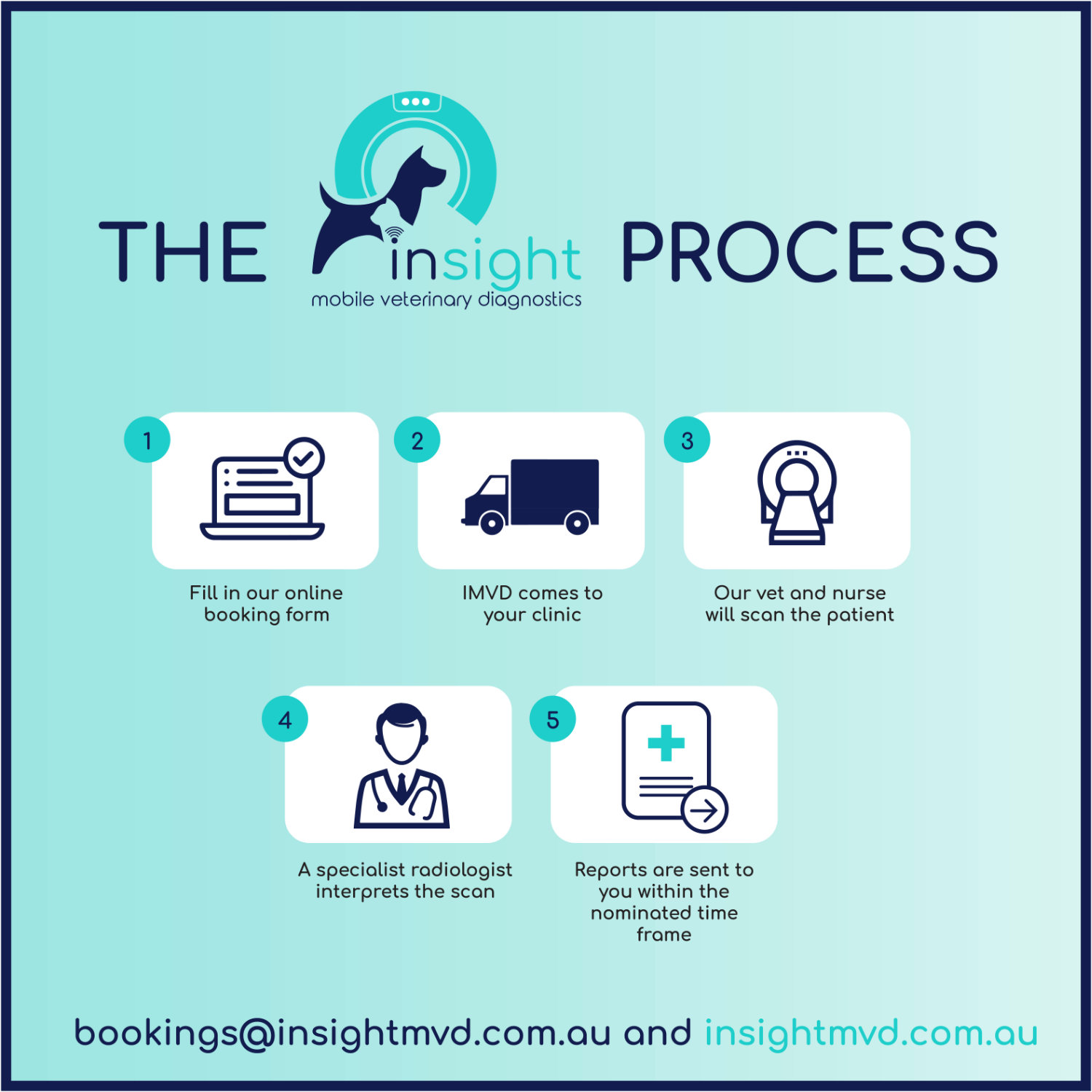
Utilising state of the art human quality Samsung NeuroLogica CT equipment, we are able to transport this to your clinic to perform on-site advanced CT scanning.
Scans are performed under general anaesthesia inside our fully equipped truck under supervision of a veterinarian and nurse.
Included in the cost of the scan is the GA, iodinated contrast, IV fluids and specialist radiologist report. As default, reports will return within 3 business days. Extra fees will apply for more urgent reports.
Book OnlineContact Us
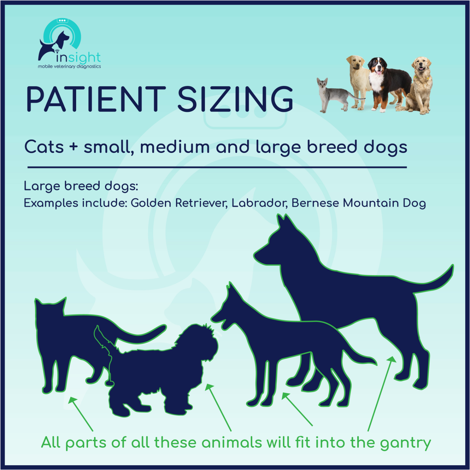
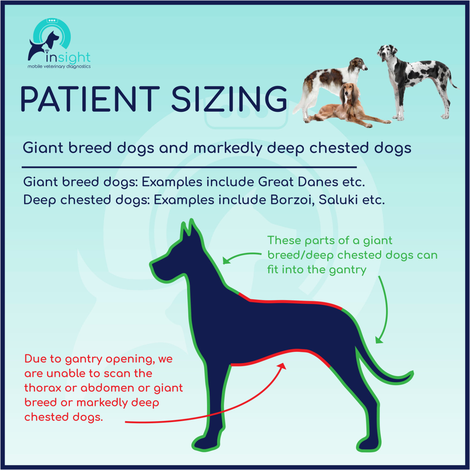
Patient Sizing
Due to the size of the gantry, we are unable to scan the thorax or abdomen or giant breed dogs (i.e Great Dane) or markedly deep chested dogs (I.e Saluki, Borzoi etc). We are still able to scan the head, neck, limbs and pelvis of these breeds.
Indications
There are so many indications about when to perform a CT scan in dogs and cats. We have summarised this and provided some information here.
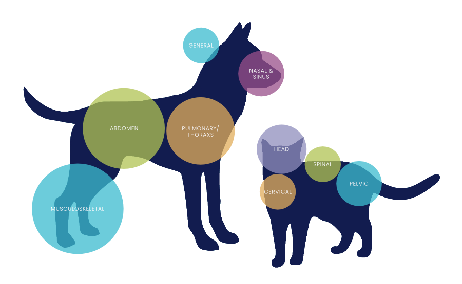
Please click each highlighted area to find out more.
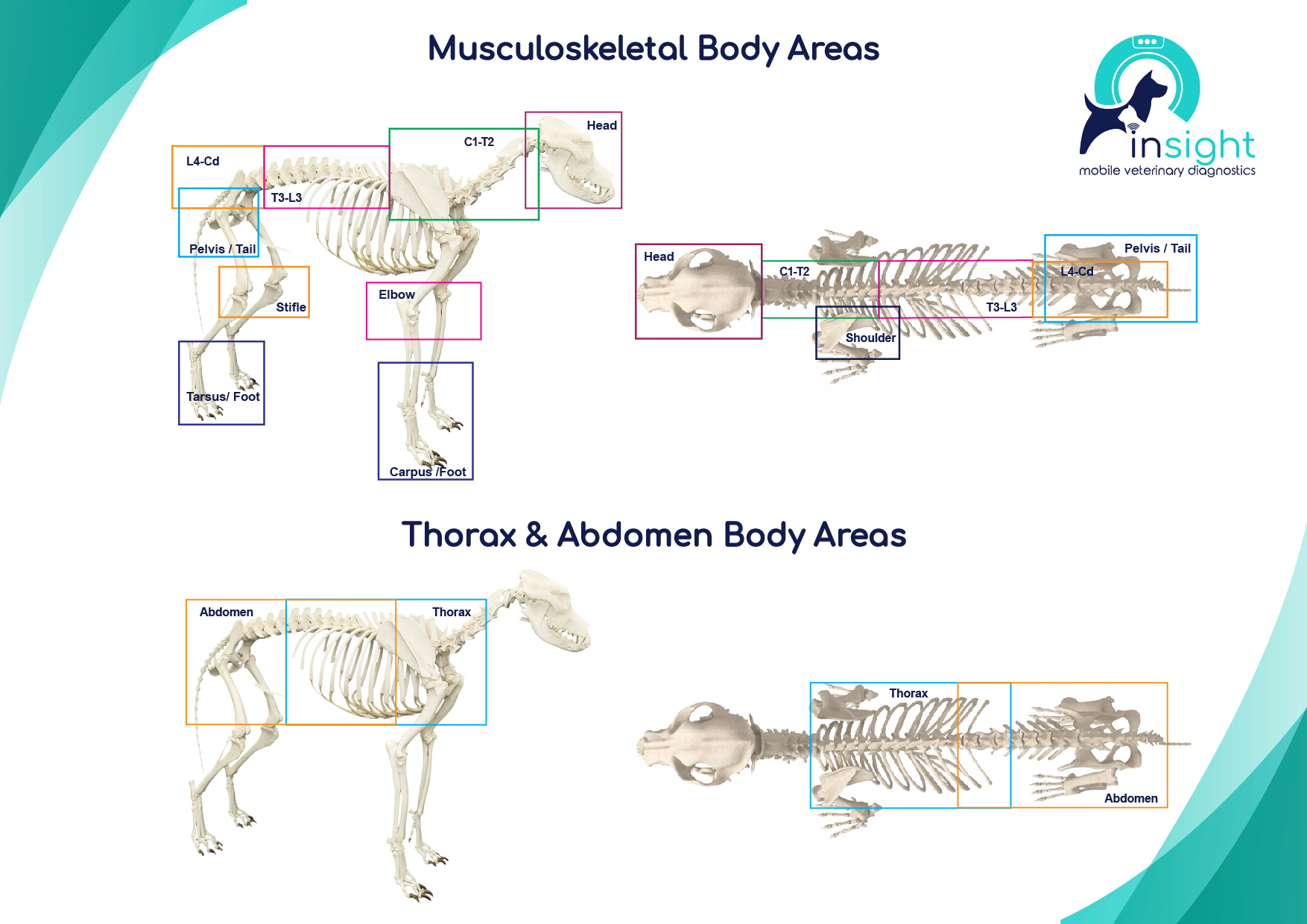
CT Scan Locations
Click the highlighted areas to view more information
Abdomen
- Evaluate organ of origin, resectability and spread for masses
- Caval or vascular invasion/thrombosis
- Ectopic ureter, ureteral disease (IVP/IVU)
- Liver shunts (CT angiography)
- Abdominal metastasis
- Organomegaly
- Diaphragmatic hernia
- Surgical planning
Head
- Any swelling/mass (eyes, mandubular, ear, etc.)
- Bulla (middle vs external ear disease distinction)
- Neurological signs of brain disease
- Chronic otitis
- Salivary gland/lymph node evaluation
- Nasopharyngeal polyp
- Head trauma
- Pituitary tumour
- Dental disease
- Stridor
- Seizures
- BOAS
- Otitis media and chronic otitis externa
- Upper respiratory sounds (stridor, snoring)
- Growths in the back of the nose, throat, and mouth
- Head trauma or injury
- Tonsils evaluation
- Neurological signs of brain disease
Cervical
- Evaluate tissue of origin, invasion, resectability and vascularity (lymph nodes, thyroid, muscle, SQ tissue) of neck masses
- Evaluate for metastasis in lymph nodes
Muscoskeletal
- Front legs: elbow dysplasia, shoulder OCD
- Back legs: stifle or tarsal OCD, hip disease
- Complicated Fractures
- Appendicular skeleton masses (muscle, SQ, bone, etc.)
- Unexplained lameness
Nasal & Sinuses
- Chronic nasal discharge
- Nasal swelling
- Epistaxis
- Nasal deformity
- Foreign body
- Fungal rhinosinusitis
- Trauma
Pelvic
- Colonic/rectal masses
- Perineal/anal gland masses
- Prostatic masses
- Urethral/vaginal masses
- Pelvic wall mass or hernia
- Perineal hernia evaluation
Spinal
- IVDD
- Spinal mass
- Trauma
- Lumbosacral disease
- Discospondylitis
- Myelopathy
- Spinal fracture
- AA luxation
Pulmonary / Thorax
- Mediastinal disease (mass, pneumomediastinum, etc.)
- Investigation into pulmonary radiographic abnormalities
- Pericardial effusion or cardiac neoplasia
- Pulmonary masses/bullae
- Pulmonary metastasis (better than x-rays)
- Pneumothorax
- Pleural effusion
- Lung lobe torsion
- Trauma (i.e., rib fracture)
- Neoplasia
- Horner's syndrome
General
- Pre-surgical evaluation of any soft tissue or osseus tumour
- Fever of unknown origin
- Migrating FB